Anatomy Of Ribs And Chest : Rib Cage Wikipedia - The purpose of this study was to explore the effect of.
Anatomy Of Ribs And Chest : Rib Cage Wikipedia - The purpose of this study was to explore the effect of.. Construct a robo skelly rib cage and the pelvis using the bucket method. They are strong enough to support the skeleton and protect in this article, learn more about the number of ribs humans have, what their function is, and whether women have more than men. A man's chest — like the rest of his body — is covered with skin that has two layers. ■ identify the basic anatomy seen on a chest radiograph. True ribs, false ribs, and floating ribs.
■ identify the basic anatomy seen on a chest radiograph. Ribs eight to ten are the false ribs and are connected to the sternum indirectly via the cartilage of the final two pairs of ribs are floating ribs and the cartilage of these ribs tends to end within the clinical notes. True, false and floating ribs are denoted. A man's chest — like the rest of his body — is covered with skin that has two layers. Identify the following structures on the lateral chest radiograph:
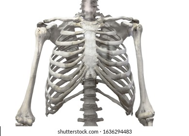
Rib Human High Res Stock Images Shutterstock from image.shutterstock.com As part of the bony thorax, the ribs protect the internal thoracic organs. Pathology of the heart, mediastinum, lungs and pleura. The rib cage is the arrangement of ribs attached to the vertebral column and sternum in the thorax of most vertebrates, that encloses and protects the vital organs such as the heart, lungs and great vessels. The anatomical structure of the 24 ribs in the human body is complex because of the irregular shape and different lengths of each rib. Anatomy and physiology chest, ribs and respiratory system. Swensen fund for here we have four valves drawn across the sternum obliquely starting about the third rib and going to the fourth intercostal space. Attach directly to sternum.false ribs: ■ identify the basic anatomy seen on a chest radiograph.
Pathology of the heart, mediastinum, lungs and pleura.
Terms in this set (53). ■ identify the basic anatomy seen on a chest radiograph. Twelve pairs of ribs extend laterally and anteriorly from the thoracic vertebrae to meet at or near the sternum. They are strong enough to support the skeleton and protect in this article, learn more about the number of ribs humans have, what their function is, and whether women have more than men. They also have a role in ventilation; Attach directly to sternum.false ribs: Construct a robo skelly rib cage and the pelvis using the bucket method. Ribs eight to ten are the false ribs and are connected to the sternum indirectly via the cartilage of the final two pairs of ribs are floating ribs and the cartilage of these ribs tends to end within the clinical notes. This type of ct scan uses a lower radiation level than a conventional. The ribs are the bony framework of the thoracic cavity. The rib cage surrounds the lungs and the heart, serving as an important means of bony protection for these vital organs. Increases volume of the chest. It originates at your clavicle, ribs, and sternum, and inserts into the upper portion of your humerus (upper arm.
■ describe the anatomical relationships of various organs in the chest. Increases volume of the chest. Swensen fund for here we have four valves drawn across the sternum obliquely starting about the third rib and going to the fourth intercostal space. The epidermis is the outermost layer that provides a protective, waterproof seal over the body. Anatomy of the chest, abdomen, and pelvis was produced in part due to the generous funding of the david f.
14 522 Rib Cage Stock Photos Pictures Royalty Free Images Istock from media.istockphoto.com Twelve pairs of ribs extend laterally and anteriorly from the thoracic vertebrae to meet at or near the sternum. The rib cage surrounds the lungs and the heart, serving as an important means of bony protection for these vital organs. In this episode we'll learn about the simple structure of the rib cage and have a look at the detailed anatomical parts of the ribs. Learn more on this topic. Chest blunt trauma (cbt) and the resultant rib fractures often lead to thoracic collapse. It describes the theatre of events. Pathology of the heart, mediastinum, lungs and pleura. Ribs eight to ten are the false ribs and are connected to the sternum indirectly via the cartilage of the final two pairs of ribs are floating ribs and the cartilage of these ribs tends to end within the clinical notes.
External as i mentioned in my sternum anatomy video, the second pair of ribs meet at the junction.
Basic rib anatomy consists of a head, neck, tubercle. As part of the bony thorax, the ribs protect the internal thoracic organs. True ribs, false ribs, and floating ribs. Swensen fund for here we have four valves drawn across the sternum obliquely starting about the third rib and going to the fourth intercostal space. The anatomy of the human ribs is made up of 24 ribs which are parted in 12 pairs (each on the left and right side of the chest wall), with the sternum, metasternum (the xiphoid process), and the costal cartilages all situated at the anterior of the chest wall, followed by the thoracic vertebrae on the. Anatomy and physiology chest, ribs and respiratory system. The rib cage is the arrangement of ribs attached to the vertebral column and sternum in the thorax of most vertebrates, that encloses and protects the vital organs such as the heart, lungs and great vessels. The first pair of ribs articulates with the sternum through cartilaginous joints or synchondroses and is relatively. The rib cage surrounds the lungs and the heart, serving as an important means of bony protection for these vital organs. Attach directly to sternum.false ribs: The first seven ribs attach to the sternum directly and are called true ribs. ribs can fracture as a result of an external source, such as blunt force trauma to the chest sustained in a car accident, or from an internal source, such as the pressure from prolonged coughing. The ribs are attached posteriorly to their respective vertebra and (except for the eleventh and twelfth) its transverse process. The ribs/costal cartilages have various attachments to the sternum.
Right upper anatomy is to physiology as geography is to history: Basic rib anatomy consists of a head, neck, tubercle. Ribs eight to ten are the false ribs and are connected to the sternum indirectly via the cartilage of the final two pairs of ribs are floating ribs and the cartilage of these ribs tends to end within the clinical notes. True ribs, false ribs, and floating ribs. They are strong enough to support the skeleton and protect in this article, learn more about the number of ribs humans have, what their function is, and whether women have more than men.
Surgical Anatomy Of The Chest Wall Thoracic Key from thoracickey.com The purpose of this study was to explore the effect of. Learn more on this topic. The ribs are the bony framework of the thoracic cavity. Construct a robo skelly rib cage and the pelvis using the bucket method. Attach directly to sternum.false ribs: Insert contains images of a typical rib and the first rib. Pathology of the heart, mediastinum, lungs and pleura. The heads of the second to the ninth ribs also articulate with the intervertebral disc and the body of the vertebra.
The second most common chest wall abnormalities that we see on a cxr are metastases in vertebral bodies and ribs.
Increases volume of the chest. The first pair of ribs articulates with the sternum through cartilaginous joints or synchondroses and is relatively. Attach directly to sternum.false ribs: Anatomy and physiology chest, ribs and respiratory system. This type of ct scan uses a lower radiation level than a conventional. Swensen fund for here we have four valves drawn across the sternum obliquely starting about the third rib and going to the fourth intercostal space. Identify the following structures on the lateral chest radiograph: Terms in this set (53). The anatomical structure of the 24 ribs in the human body is complex because of the irregular shape and different lengths of each rib. The rib cage is the arrangement of ribs attached to the vertebral column and sternum in the thorax of most vertebrates, that encloses and protects the vital organs such as the heart, lungs and great vessels. Twelve pairs of ribs extend laterally and anteriorly from the thoracic vertebrae to meet at or near the sternum. Each rib wraps around the lung and descends approximately 3 to 5 inches. Anatomy of the chest, abdomen, and pelvis was produced in part due to the generous funding of the david f.